File:Human karyotype with bands and sub-bands.png

From Wiktionary, the free dictionary
Jump to navigation Jump to search

Original file (9,684 × 16,008 pixels, file size: 9.58 MB, MIME type: image/png)

Any autoconfirmed user can overwrite this file from the same source. Please ensure that overwrites comply with the guideline.

Summary

Warning The original file is very high-resolution. It might not load properly or could cause your browser to freeze when opened at full size. Open in ZoomViewer
Description

Add language/edit

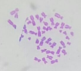
English: Schematic and relatively language-neutral karyogram of a human. Even at low magnification, it gives an overview of the human genome, with numbered chromosome pairs, its main changes during the cell cycle (top center), and the mitochondrial genome to scale (at bottom left). High magnification (of full size version) more clearly depicts a scale to the left of each chromosome pair that shows the length in terms of million base pairs, and the scale to the right of each chromosome pair shows the designations of the bands and sub-bands. It shows darker and brighter regions as seen on G banding. Each row is vertically aligned at centromere level. The chromosomes are organized into the groups A to G in regard to size, position of the centromere and sometimes the presence of a chromosomal satellite. The chromosome copy numbers are those seen in G0 and G1 of the cell cycle, and at top center it also shows the state of the chromosome 3 pair in the metaphase of the cell cycle, after the synthesis of a copy of each chromosome. The karyogram shows:
- 22 homologous autosomal chromosome pairs
- Both the female (XX) and male (XY) versions of the two sex chromosomes (in green rectangle at bottom right).
- The human mitochondrial genome to scale (at bottom left).
  • Printing: For a regular printer, adjust to fill at least 2 x 2 pages (A4 or standard letter size) in portrait mode, and preferably print in color on photo paper. For poster printing, the suggested size is approximately 47.0 x 77.7 cm (18.5 x 30.6 inches). The pdf version is 20 x 32 inches.

  • Svenska: Schematiskt och relativt språkneutralt karyogram av människan. Även på låg förstoring ger den en översikt över det mänskliga genomet, med numrerade kromosompar, deras huvudsakliga förändringar under cellcykeln (överst i mitten), och mitokondriernas genom i samma skala (längst ner till vänster). Högre förstoring (av fullstorleksversionen) ger tydligare en skala till vänster om varje kromosompar som visar längden i form av miljoner baspar, och skalan till höger om varje kromosompar visar beteckningarna för band och sub-band. Den visar mörkare och ljusare områden som ses på G-banding (med Giemsa-färjning). Varje rad av kromosomer är justerat så att varje centromer ligger i samma nivå. Kromosomerna är organiserade i grupperna A till G med avseende på storlek, placering av centromeren och ibland närvaron av en kromosomsatellit. Antalet kromosomkopior är de som ses i G0 och G1 i cellcykeln. Karyogrammet visar:
    - 22 homologa autosomala kromosompar
    - Både den kvinnliga (XX) och den manliga (XY) versionen av de två könskromosomerna (i grön rektangel längst ner till höger).
    - Det mänskliga mitokondriella genomet i samma skala (längst ner till vänster).
    • Mer detaljerad beskrivning av detta karyogram finns på engelska Wikipedia: Wikipedia:Karyotype.
  • Utskrift: För en vanlig skrivare, justera till att fylla minst 2 x 2 sidor (A4) i stående läge. För affischtryck är en föreslagna storleken cirka 47.0 x 77.7 cm, och helst i färg på fotopapper. pdf-versionen är 20 x 32 tum (50.8 x 81.28 cm).
  • العربية: رسم بياني للشكل الخلوي المضاعف للصبغيات عند الإنسان، يبين تنظيم الجينوم البشري على هيئة صبغيات حتى قبل تكبير الصورة. يُظهر الرسم أزواج الصبغبات مرقمة، والتغييرات الرئيسة خلال دورة الخلية (في الأعلى والمركز)و علم الوراثة البشري الميتوكوندري (في الأسفل على اليسار). تصور تصويراً واضحاً الصورة بالدقة العالية المقياس الأيسر طول الأزواج الصبغية مقدراً بالمليون، فيما يظهر أيمن الزوجين الصبغيين تسميات العصابات والنطاقات الفرعية. يظهر مناطق أكثر قتامة وإشراقا كما يظهر في النطاق G. يتم محاذاة كل صف عموديا عند مستوى سنترومير. يتم تنظيم الكروموسومات في المجموعات من أ إلى ز فيما يتعلق بحجم وموقع السنترومير وأحيانا وجود قمر صناعي كروموسومي. أرقام نسخ الكروموسوم هي تلك التي تظهر في G0, G1 من دورة الخلية، وفي المركز العلوي يظهر أيضا حالة زوج الكروموسوم 3 في الطور لدورة الخلية ، بعد تخليق نسخة من كل كروموسوم. يظهر مخطط كاريوجرام:22 متماثل جسمي أزواج كروموسوم - كلا النسختين الأنثوية (س س) والذكور (س ع) من الاثنين كروموسوم جنسي (في مستطيل أخضر في أسفل اليمين).- الجينوم الميتوكوندريا البشرية لتوسيع نطاق (في أسفل اليسار).


    中文:相对语言中性的人类核型图示意图。即使在低倍放大下,它也能概览人类基因组,包括编号的染色体对,细胞周期(顶部中心)中的主要变化,以及按比例显示的线粒体基因组(左下角)。高倍放大(完整版本)更清晰地显示了每对染色体左侧的刻度,表示以百万碱基对为单位的长度,右侧的刻度显示了条带和亚条带的标记。它显示了在G显带上看到的深浅不同的区域。每一行在着丝粒水平垂直对齐。染色体根据大小、着丝粒位置以及有时是否存在染色体卫星被组织为A到G组。染色体的拷贝数是在细胞周期的G0和G1期看到的,顶部中心还显示了细胞周期中期时第3对染色体的状态,即每条染色体复制一份后的状态。核型图显示:
    - 22对同源常染色体
    - 两种性染色体的女性(XX)和男性(XY)版本(右下角绿色矩形中)。
    - 按比例显示的人类线粒体基因组(左下角)。
    • 更多信息和背景,请参见zh:核型
  • 打印: 对于普通打印机,调整以填满至少2 x 2页(A4或标准信纸大小)的纵向模式,最好在照片纸上彩色打印。对于海报打印,建议尺寸约为47.0 x 77.7厘米(18.5 x 30.6英寸)。PDF版本尺寸为20 x 32英寸。

    Add language/edit
  • Date
    Source

    edit
    Own work, integrating the following Public Domain pictures:

    Animal Cell.svg by Kelvin Ma (for mitochondrial localization)

    The purple hue for the chromosomes and cell nuclei was digitally sampled from the following image:

    Author
    Mikael Häggström, M.D. Author info
    - Reusing images
    - Conflicts of interest:
      None
    Mikael Häggström, M.D.
    - The work integrates Public Domain source images by Was A Bee, Kelvin Ma and Michał Komorniczak
    - Also, a special acknowledgement to everyone involved in the Human Genome Project for generating the source data.
    edit
    Other versions

    edit

    Assessment

    Featured picture

    Wikimedia CommonsWikipedia

    This is a featured picture on Wikimedia Commons (Featured pictures) and is considered one of the finest images. See its nomination here.

     This is a featured picture on the Arabic language Wikipedia (صور مختارة) and is considered one of the finest images. See its nomination here.
     This is a featured picture on the English language Wikipedia (Featured pictures) and is considered one of the finest images. See its nomination here.

    If you have an image of similar quality that can be published under a suitable copyright license, be sure to upload it, tag it, and nominate it.

    Licensing

    Creative Commons CC-Zero This file is made available under the Creative Commons CC0 1.0 Universal Public Domain Dedication.
    The person who associated a work with this deed has dedicated the work to the public domain by waiving all of their rights to the work worldwide under copyright law, including all related and neighboring rights, to the extent allowed by law. You can copy, modify, distribute and perform the work, even for commercial purposes, all without asking permission.

    Captions

    Schematic karyogram of a human. Even at low magnification, it gives an overview of the human genome, with numbered chromosome pairs, its main changes during the cell cycle (top center) and the mitochondrial genome to scale (at bottom left).

    Items portrayed in this file

    depicts

    20 November 2022

    image/png

    File history

    Click on a date/time to view the file as it appeared at that time.

    (newest | oldest) View (newer 10 | ) (10 | 20 | 50 | 100 | 250 | 500)
    Date/TimeThumbnailDimensionsUserComment
    current14:59, 20 April 2024Thumbnail for version as of 14:59, 20 April 20249,684 × 16,008 (9.58 MB)Obscure2020The file was previously encoded as RGBA, which unnecessarily inflated the file size, because the image is completely opqaue. I have corrected the image to RGB with the help of OxiPNG.
    03:43, 7 February 2023Thumbnail for version as of 03:43, 7 February 20239,684 × 16,008 (11.83 MB)Mikael HäggströmMinor adjustments
    01:06, 6 February 2023Thumbnail for version as of 01:06, 6 February 20239,684 × 16,008 (11.83 MB)Mikael HäggströmRemoved excessive margins
    01:03, 6 February 2023Thumbnail for version as of 01:03, 6 February 202311,600 × 17,789 (12.1 MB)Mikael HäggströmTiny adjustment
    02:26, 5 February 2023Thumbnail for version as of 02:26, 5 February 20239,684 × 16,008 (11.83 MB)Mikael HäggströmSlight reduction in size without affecting relevant information
    04:04, 9 January 2023Thumbnail for version as of 04:04, 9 January 202313,887 × 22,956 (18.88 MB)Mikael HäggströmSame gray hue
    03:51, 9 January 2023Thumbnail for version as of 03:51, 9 January 202313,887 × 22,956 (18.88 MB)Mikael HäggströmSwitched to grey around chromosomes and white around. Some other adjustments
    04:11, 30 December 2022Thumbnail for version as of 04:11, 30 December 202213,887 × 22,956 (20.04 MB)Mikael HäggströmMinor adjustments
    03:40, 28 December 2022Thumbnail for version as of 03:40, 28 December 202213,887 × 22,956 (20.04 MB)Mikael HäggströmImproved metaphase cell
    18:29, 24 December 2022Thumbnail for version as of 18:29, 24 December 202213,887 × 22,956 (20.02 MB)Mikael HäggströmMore realistic shapes of metaphase cell and chromosomes
    (newest | oldest) View (newer 10 | ) (10 | 20 | 50 | 100 | 250 | 500)

    The following page uses this file:

    Global file usage

    The following other wikis use this file:

    View more global usage of this file.

    Metadata